You are using an out of date browser. It may not display this or other websites correctly.
You should upgrade or use an alternative browser.
You should upgrade or use an alternative browser.
Ring Bone
- شروع کننده موضوع د.شهبازی
- تاریخ شروع
امین سلطانی
Member
کسی در مورد ring bone نظری ،اطلاعاتی نداره؟دکتر جان فکر نکنم یه عکس کفایت کنه...
Ringbone is a lay term used to describe an osteoarthritis that affects the pastern joints in both the front and hind limbs of horses. Simply put, ringbone is new bone growth on the proximal, middle, or distal phalanx often with degeneration of the joint surface. High ringbone is the term that has been applied to the condition when it affects the proximal interphalangeal joint (pastern joint). Low ringbone is the term that has been applied when the condition affects the distal interphalangeal joint (coffin joint). Please refer to figure 1 for a brief review of the structures of the foot and pastern region.

[FONT=Arial, Helvetica, sans-serif][SIZE=-1]A. First Phalanx [/SIZE][/FONT]
[FONT=Arial, Helvetica, sans-serif][SIZE=-1]B. Proximal interphalangeal joint (pastern joint) [/SIZE][/FONT]
[FONT=Arial, Helvetica, sans-serif][SIZE=-1]C. Second Phalanx [/SIZE][/FONT]
[FONT=Arial, Helvetica, sans-serif][SIZE=-1]D. Distal interphalangeal joint (coffin joint) [/SIZE][/FONT]
[FONT=Arial, Helvetica, sans-serif][SIZE=-1]E. Third Phalanx [/SIZE][/FONT]
[FONT=Arial, Helvetica, sans-serif][SIZE=-1]Figure 1[/SIZE][/FONT]
Since ringbone is a disorder affecting the proximal and distal interphalangeal joints, we should briefly review the anatomy of a typical synovial joint. A synovial joint has two major functions. The first is to enable movement and the second is to transfer load. Therefore the structure of a joint is designed to facilitate these functions. The proximal interphalangeal joint is considered to be a low motion joint and contributes only a small percentage to the flexion or extension of the foot/pastern region.
A typical synovial joint is comprised of two bones articulating one withanother. The articular surface of the two bones is covered with a type ofcartilage called hyaline cartilage. The space between the two bones is filledwith a viscous fluid that both lubricates the joint and provides nutrition tothe cells within the cartilage. This fluid is secreted by cells that comprisethe synovial lining. A strong fibrous joint capsule surrounds the joint andinserts on the adjacent bone a variable distance from the articulating jointsurface. Finally, strong fibrous collateral ligaments are attached to the boneon each side of the joint and lie closely associated on the outside of thejoint capsule. Their function is to provide medial and lateral stability to thejoint.
Ringbone has been described as being articular or periarticular. Articularringbone exists when there is radiographic evidence of degenerative jointdisease such as partial or complete narrowing of the joint space, new boneformation at the joint margins (osteophytes), increased density of thesubchondral bone (sclerosis), or subchondral bone erosion (lysis).Periarticular ringbone exists when there is radiographic evidence of new bonegrowth away from the joint margins. This new bone growth can occur at sites ofligament or joint capsule origins or insertions (enthesophytes). New bonegrowth can also occur anywhere the connective tissue that covers the bone (theperiosteum) may have been damaged by external trauma such as blunt trauma ordeep cuts. Figure 2 displays normal joint structures on the left anddegenerative changes on the right.
[FONT=Arial, Helvetica, sans-serif][SIZE=-1]A. Periosteum [/SIZE][/FONT]
[FONT=Arial, Helvetica, sans-serif][SIZE=-1]B. Collateral Ligament [/SIZE][/FONT]
[FONT=Arial, Helvetica, sans-serif][SIZE=-1]C. Joint Capsule [/SIZE][/FONT]
[FONT=Arial, Helvetica, sans-serif][SIZE=-1]D. Subchondral bone [/SIZE][/FONT]
[FONT=Arial, Helvetica, sans-serif][SIZE=-1]E. Synovial Space narroving joint space, erosion channel
[/SIZE][/FONT]
[FONT=Arial, Helvetica, sans-serif][SIZE=-1]F. Articlular hyaline cartilage [/SIZE][/FONT]

[FONT=Arial, Helvetica, sans-serif][SIZE=-1]1. Periosteal Elevation [/SIZE][/FONT]
[FONT=Arial, Helvetica, sans-serif][SIZE=-1]2. Capsulitis [/SIZE][/FONT]
[FONT=Arial, Helvetica, sans-serif][SIZE=-1]3. Osteophyte [/SIZE][/FONT]
[FONT=Arial, Helvetica, sans-serif][SIZE=-1]4. Carilage necrosis, [/SIZE][/FONT]
[FONT=Arial, Helvetica, sans-serif][SIZE=-1]5. Enthesophyte [/SIZE][/FONT]
[FONT=Arial, Helvetica, sans-serif][SIZE=-1]Figure 2[/SIZE][/FONT]
Several factors contribute to ringbone. These are generally grouped into conformational defects, traumatic insults, or developmental factors. Poor conformation has been identified as being a major contributing factor to the development of ringbone. Pasterns that are overly upright contribute to increased concussion of the pastern joint. Angular limb deformities and horses that may toe-in or toe-out distribute abnormal stresses across the joint surfaces as well as on the joint capsule and ligaments.
Trauma induced ringbone can occur following strain-sprain type injuries to thesoft tissue support structures of the joint. In fact, ringbone occurs morecommonly in horses that make sharp turns and sudden stops such as westernperformance horses and polo ponies. A fracture that involves the joint surfaceor the extensor process of the third phalanx, and luxations or subluxations ofthe joint can all lead to the formation of ringbone. Joint infections resultingfrom wounds that penetrate into the joint can also ultimately lead to ringbone.
A syndrome has also been described in young horses in which lesions ofosteochondrosis or failure of the normal developmental process by whichcartilage is changed to bone, are seen in conjunction with those ofdegenerative joint disease in the proximal interphalangeal joint.
Movement is essential for maintaining the normal physiologic processes,structure, and mechanical function in synovial joints. The cells of articularcartilage are metabolically active. The movement of water, soluble nutrients,and wastes within the cartilage matrix is facilitated when the articularcartilage is cyclically loaded during locomotion. For this reason, low-motionjoints such as the proximal interphalangeal joint are especially vulnerable tothe development of osteoarthritis if their already limited range of motion isfurther restricted. A proposed mechanism for the development of osteoarthritisin these joints is that trauma to the periarticular soft tissue surrounding thejoint results in pain, inflammation, edema, fibrosis of the joint capsule,enthesophyte formation, and periosteal new bone formation at the joint capsuleinsertion. The pain and the changes in the soft tissue reduce the alreadylimited range of motion within the joint and therefore the weight bearingforces across the joint are now focused on more focal areas of the jointsurface. This sustained compression on focal areas of cartilage and thesupporting bone beneath it leads to the destruction of the cartilage andstimulates the bone to remodel. In some cases, the remodeling bone actuallybridges across the joint surface in an attempt to fuse the joint together.
Ringbone may occur in either the front limbs or the hind limbs, but isgenerally more common in the front limbs and often occurs in both front limbsat the same time. The initial signs are generally not specific and may rangefrom a decrease in the normal performance to a variable progressive lameness.In cases where fracture or infections are the inciting cause, the lameness isgenerally severe and acute in onset. With osteochondrosis lesions in younghorses, there is usually a progressive lameness and marked pastern swelling.

[FONT=Arial, Helvetica, sans-serif][SIZE=-1]A. Enlarged proximal interphalangeal joint [/SIZE][/FONT]
[FONT=Arial, Helvetica, sans-serif][SIZE=-1]Figure 3[/SIZE][/FONT]
[FONT=Arial, Helvetica, sans-serif][SIZE=-1]Figure 3[/SIZE][/FONT]
In the early stages, especially with use related trauma, there may be a transient warm swelling in the pastern region that resolves with rest. Affected horses may show sensitivity to pressure over the pastern region and flexion of the pastern may exacerbate the lameness. As the condition becomes more chronic the typical outward appearance of ringbone becomes evident with firm, cool swelling around the pastern joint. Figure 3 depicts the typical appearance of high ringbone. These horses often are more progressively lame with a decreased range of motion in the pastern region. Some cases of ringbone can be relatively asymptomatic especially if the new bone growth is periarticular. In contrast, some early cases may have soft tissue swelling but do not yet have radiographic changes in the bone. In these cases further radiographs are often taken several weeks later to evaluate any new bone production.
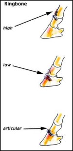
The use of diagnostic nerve blocks and intra-articular anesthesia is oftennecessary to determine whether the suspected structures are in-fact the causeof the lameness. In any case, radiographs are necessary for a definitivediagnosis of ringbone. Figure 4 depicts some of the radiographic findings withhigh ringbone.
Before we discuss the treatment I think is important to keep in mind thatringbone is generally a progressive condition that will continue to producelameness, especially if there is evidence of degenerative joint disease. Somecases, especially those with periarticular changes, can be managed effectivelywith athletic soundness for several years.
The treatment of ringbone is directed by the inciting cause, the duration ofthe condition, the degree of lameness, and the intended use of the horse. Theprimary goal should be to minimize the factors that contribute to itsdevelopment. This should include a critical evaluation of the conformation of ahorse and its suitability for your intended use. If you are consideringpurchasing a horse it is a good idea to have a veterinarian perform apre-purchase exam and possibly take radiographs of the pastern region ifproblems are suspected. If you already own a horse that has a conformation thatwould contribute to abnormal forces across these joints, then regular scheduledhoof care is important. This includes the proper balancing of the foot andappropriate shoeing. Finally, take the time to adequately warm-up a horsebefore competition or any strenuous activity.
If your horse shows a sudden onset of lameness, it is important to have itevaluated by a veterinarian to determine the cause. In the case of astrain/sprain type of injury to the joint your veterinarian may prescribe anumber of treatment options directed at reducing the pain and inflammation.These may include rest, anti-inflammatory drugs, leg wraps and hot or coldtherapy.

[FONT=Arial, Helvetica, sans-serif][SIZE=-1]A. Radiographic appearances of degenerative changes in proximal interphalangeal joint [/SIZE][/FONT]
[FONT=Arial, Helvetica, sans-serif][SIZE=-1]Figure 4[/SIZE][/FONT]
[FONT=Arial, Helvetica, sans-serif][SIZE=-1]Figure 4[/SIZE][/FONT]
If there is evidence that the condition is more chronic in nature it is important to keep in mind that this does tend to be a progressive condition. The initial course of treatment for chronic cases is usually attempted by treating the condition medically.
Medical management of these chronic cases may include the judicious use ofnon-steroidal anti-inflammatory drugs, intra-articular corticosteroidinjections and appropriate shoeing with a roller motion shoe that willfacilitate easier break over of the foot. Often medical management may extendthe athletic career of horses for a variable duration. In many cases however,medical management alone will not result in long term soundness and the onlyoption remains surgical intervention.
Surgical treatment involves fusing the pastern joint to prevent joint movementand thus prevent further pain. Remember the previous discussion of thepathogenesis of the condition where often the articular cartilage is destroyedand the bone begins to bridge across the joint. Surgical intervention seeks toaccelerate this process by removing the articular cartilage and fusing thejoint with screws and plates. The limb is then placed in a half limb cast untilthe joint is adequately fused. The goal of this procedure is to produce a soundhorse. Published success rates have been from 67% for the front limbs and 75%to 80% for the hind limbs. Some surgeons report even higher success rates basedupon their clinical experience. It is generally accepted that the prognosis islower for the front limbs than the hind limbs.
Ringbone is a progressive, degenerative, performance limiting condition. It ishowever one of many possible causes of lameness that occurs in the lower limbof the horse. Many of these conditions also produce very similar initial signs.For this reason it is important to have your horse evaluated by yourveterinarian in order to accurately diagnose, treat, and provide a prognosisfor the continued soundness of your horse.
