Anatomy and Topography of the Equine Foot

♘امیرحسین♞

♘ مدیریت انجمن اسب ایران ♞
The modern horse would have little value other than for its possible breeding potential without healthy feet. Nevertheless, the foot has been the most neglected part of the horse since its early domestication. Close confinement, exaggerated gaits produced by intensive training, and poor conformation as a result of man's selective breeding for other purposes, have all compounded this problem. We hope that this discussion will help horsemen understand hoof functions and will provide standard names for parts that have been called by many different names in the past.

Topography of the Hoof
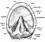
This description of the anatomy of the foot entails the hoof and all of the structures within it, with some reference to certain related structures. The hoof itself is nothing more than cornified epidermis , similar in makeup to man's fingernail, and contains no nerves or blood vessels but relies upon the corium , another inner layer just interior to the white line, to provide the circulation and sensitivity necessary to maintain a healthy foot. Since the hoof itself is not sensitive, we must understand its relationship to the inner structures in order to comprehend any lameness related to the hoof. The nomenclature (set of names) of the external parts of the hoof is divided into the following areas ( Figure 1 ):​
 

پیوست ها

  • Figure 1.gif
    Figure 1.gif
    18.4 کیلوبایت · بازدیدها: 0

♘امیرحسین♞

♘ مدیریت انجمن اسب ایران ♞
1. The bulbs of the heel . These structures are at the back part of the ground surface of the foot, behind the angle of the hoof wall. Internally they receive support from the digital (plantar) cushion .
2. The frog . This is a triangular or wedge-shaped structure with the apex pointing toward the front of the hoof and is terminated by the base at the rear. Two grooves, one on each side of this structure are known as the collateral sulcus . The central ridge is named the frog stay or spine of the frog and contains a furrow called the central sulcus or cleft .
3. The bars . Near the heel the wall of the hoof turns back toward the front portion and this continuation forms a ridge on both sides of the frog known as the bars .
4. The wall . This surrounding structure encompasses the foot and all inner parts. It is elastic in nature and continually grows downward from the coronet. It is divided into three general areas: the toe, quarter , and the heel .
5. The sole . The sole makes up a major portion of the surface area of the bottom of the hoof, but is not designed to bear the weight of the animal. It provides support for the internal structures.
6. The white line . This boundary serves as a junction between the wall and the sole and is clearly visible around the front three-fourths of the circumference of the sole.

The Internal Anatomy of the Foot
Describing the structure of the inside of the horse's foot is complicated by the relationships between the different parts and by the different types of body tissue that make up these parts. Also, different names have often been used for the same structures in both technical literature and in everyday language.

Figure 2 shows some of the duplication of the nomenclature and these duplications will also be listed in the text. This should eliminate some of the confusion in this area.​
 

پیوست ها

  • Figure 2.gif
    Figure 2.gif
    14.4 کیلوبایت · بازدیدها: 1

♘امیرحسین♞

♘ مدیریت انجمن اسب ایران ♞
Bones of the Foot

1. Third phalanx (P 3 ) is also called the distal phalanx, os pedis, pedal bone , and the coffin bone . It is the most distal (farthest out from the body) of the four bones comprising the digit (equivalent to man's finger or toe) and is completely enclosed by the hoof. Interaction between this bone and the surrounding hoof structures serves as a shock absorber for the horse in motion.
2. The Second Phalanx (P 2 ) is also referred to as the middle phalanx, os coronale , and the short pastern bone . This bone rests on the 3rd phalanx and articulates with it and the lst phalanx which is above it.
3. The Distal Sesamoid , commonly called the navicular bone or shuttle bone is located on the back surface of both the 2nd and 3rd phalanx and is an integral part of the anticoncussive (shock absorbing) mechanism along with its ligamentous attachments.
4. The First Phalanx (P 1 ) as with the other bones of the digit has its synonyms: os compendale, os saffragenous , and the long pastern bone . The lst phalanx is the longest bone of the digit, rests on the 2nd phalanx and also articulates with the 3rd metacarpal or 3rd metatarsal depending upon whether its location is the fore leg or hind leg. It is closely attached to the paired proximal sesamoids by strong ligaments.

Other Structures Within the Foot

1. The corium or pododerm can be divided into five parts, but for our discussion we will consider only the general term and its function as a nutritional source to the hoof. Within this structure lies a massive supply of blood vessels that feed the hoof. These blood vessels combined with nerves form a sensitive layer intimately attached to the inside of the hoof wall and the 3rd phalanx .
2. The digital (plantar) cushion is a wedge-shaped modified subcutaneous tissue located within the back part of the hoof and is composed of elastic fibers and some cartilage. As the name implies, it serves to reduce concussion to the foot.
3. The tendon of the common digital extensor muscle is considered here because of its insertion as a process of the 3rd phalanx and on the anterior (front) surfaces of the 2nd and lst phalanges . Its action is to extend the entire digit.
4. The deep flexor tendon is an extension of the muscle lying on the back part of the leg and inserting on the posterior aspect of the 3rd phalanx .
5. The superficial flexor tendon runs parallel to the deep flexor tendon and divides below the fetlock and inserts on both the lst and 2nd phalanges .

Conclusion
With a better understanding of the anatomy of the hoof gained from this discussion we hope that better hoof care will result. The information presented here should be the basis for understanding why a horse must be shod or have its feet trimmed on a regular basis and provide some insight as to how some of the more common injuries affect the hoof and the way a horse travels. In turn this understanding should also help to prevent injuries to the hoof. A horse that has healthy feet is the horse that can provide satisfaction and pleasure to its owner.​
 
بالا