www.newenglandequine.com
Melanoma in grey horses is an often benign neoplasm (cancer) of melanocytes (pigment cells) that is usually found in the skin, although it can be found in other organs. [SUP]3[/SUP] It is one of the most prevalent neoplasias seen, and it is described to be nearly ubiquitous in 8 to 10 year old grey horses, whereas it is much less common in horses of other colors. [SUP]3[/SUP] One study estimates that up to 80% of grey horses over 15 years old develop melanoma. [SUP]4[/SUP] Melanomas in grey horses are most often non-invasive. [SUP]3[/SUP]Unlike human malignant melanoma, grey horse melanomas are encapsulated, and metastatic progression is prevented or slowed down by factors that are not yet known. [SUP]4[/SUP]
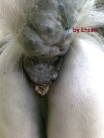
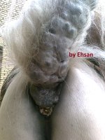
The tumors can ulcerate and release a dark tarry discharge. [SUP]3[/SUP] One author described that spherical to ovoid, tightly covered, shining black lesions in the predilection sites of grey horses greater than 7-9 years are pathognomonic for the condition.” [SUP]3[/SUP]
When someone is speaking about equine melanoma, they will often be referring to the slow growing, often benign melanomas of older grey horses that frequently cause no problem to horse or owner. This is because these are much more common than the alternative. But to be complete, it is important to know that there are four classifications of melanoma in equids.
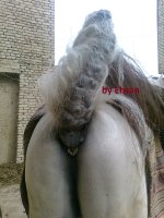
1. Benign Dermal Melanoma[SUP]4, 5[/SUP]: These are the tumors that develop in older grey horses, frequently under the base of the tail, in the anus, perineum or genitalia, perineum, lips, or eyelids. These tend to be nodules that rarely metastasize. Most common.
2. Malignant Dermal Melanomatosis[SUP]4, 5[/SUP]: This describes a state of multiple interspersed dermal melanomas that have a high likelihood of metastasizing. This occurs in grey horses, often those that are greater than 15 years old. Like benign dermal melanomas, these tend to occur at the base of the tail, in the perineum, near the genitalia and near the anus.
3. Melanocytic nevi[SUP]4, 5[/SUP]: These are seen in younger horses irrespective of color (both grey and non-grey horses) and they are superficial, benign and discrete lesions.
4. Anaplastic malignant melanoma[SUP]4, 5[/SUP]: These are very rarely seen, they tend to occur in non-grey horses that are older, and they tend to cause organ metastasis (spread to other body organs) that eventually leads to death.
Benign dermal melanomas and malignant dermal melanomatosis look very similar on histopathology, and they both occur in grey horses in the same predilected regions. [SUP]4[/SUP] Therefore, neither the location of the tumor nor the histology can help you to predict the tumor’s potential
to form a melanomatosis. One helpful factor that can help to determine the behavior of a tumor is its rate of growth.[SUP] 2 [/SUP] A rapid change in growth rate has been linked to malignant transformation.[SUP]2[/SUP] According to Pulley and Stannard, the most common sites for internal organ metastasis are regional lymph nodes and lungs. Other possible sites include the spleen, liver, blood vessels and the heart.[SUP]2[/SUP]
The etiology of melanomas in horses has not been clearly determined. One dominant theory is that melanomas in older grey horses are due to disturbed metabolism of melanin. This is thought to lead to the development of new melanoblasts, or possibly to increased activity of melanoblasts in the area resulting in a central area of too much pigment production in the dermis. It is controversial as to whether ultraviolet radiation (sun exposure) plays a role in equine melanoma
as it does in humans. Because most melanoma lesions occur in regions not exposed to the sun, it seems unlikely that the sun plays as significant a role as it does in human melanoma.[SUP]6[/SUP]
When grey horses are born, they have dark hair coats and skin. [SUP]4[/SUP] As they grow older, their hair turns white while their skin remains dark grey in most cases. In some grey horses, their skin also becomes lighter as they age. People theorize that the high prevalence of melanoma in grey horses is due to the changes that occur in the skin and hair coat pigmentation with aging.[SUP]4[/SUP] Some theorize that equine melanoma may represent a storage disease rather than a true cancer.[SUP]3[/SUP]
However, another study specifically stated their findings that grey and non-grey horse melanomas are actual neoplasms and they resemble human malignant melanomas in terms of histological traits and immunostaining.[SUP]4[/SUP]
The most common locations for equine melanoma are the groin, the perineal skin, the lips, and the eyelids. [SUP]3 [/SUP] Other common locations include the parotid lymph nodes and salivary glands.[SUP] 3[/SUP] Clinical signs depend on the location and size of the tumor.
To diagnose melanoma, biopsy is rarely necessary. Fine needle aspirate of a lesion is usually sufficient because it will yield melanin-producing cells (melanocytes).[SUP]3[/SUP] Other differential diagnoses include equine sarcoid, fibroma, eosinophilic granulomas, neoplasia or infection of the salivary glands, lymph node abscessation, and melanosarcoma. [SUP]3[/SUP]
To fully evaluate the extent of a horse’s melanoma, diagnostics would include rectal palpation, ultrasonography, biopsy or fine needle aspirate of the mass, and cytology of the abdominal fluid. In some cases, examination of the guttural pouch can be diagnostic for melanoma. On guttural pouch endoscopic exam, melanomas will appear like distinct, spherical, dense black masses. [SUP]3[/SUP] Melanoma is more common in the lateral compartment of the guttural pouch and over the
maxillary artery than in the medial compartment of the guttural pouch.[SUP]3[/SUP]
Often, veterinarians do not pursue treatment of melanoma in horses because they are believed to be benign and slow growing, because the lesions have become too large, because the lesions are close to major blood vessels or important structures, or because there has been extensive invasion into local tissues.[SUP] 2[/SUP] One study states that all dermal melanomas should be considered as possibly malignant and treated as such.[SUP]2[/SUP]
A single isolated lesion may not cause problems on its own initially, but should be removed because it will likely grow larger, and early excision prevents later ulceration and complications associated with larger size. [SUP]3[/SUP] It is harder to do a large radical excision than to do an early small incision.[SUP] 3[/SUP] Unlike with equine sarcoid, early surgery does not worsen the malignancy with melanoma. [SUP]3[/SUP] Excision of the melanocytic nevi and the benign dermal melanomas has been described to be curative. [SUP]5[/SUP]
Oral cimetidine (3.5 mg/kg BID), an agonist of the histamine receptor, for up to 3 months reportedly reduces growth of the tumor, as shown in a few cases. [SUP]3[/SUP] Histamine stimulates T suppressor cells to inhibit cell-mediated and humoral immunity. Cimetidine blocks histamine
receptors on these cells, increasing the body’s ability to fight the tumor with the immune system.
Treatment options include (1) surgical excision of isolated lesions (not possible with large or multiple confluent masses), (2) cryosurgery followed by surgical excision of the parts of tumor that are most accessible (low success rate reported), and (3) intralesional cisplatin (injection of cisplatin emulsion, or placement of cisplatin-containing biodegradable beads).
Intra-lesional cisplatin has been used frequently to treat cutaneous neoplasias in horses and has been effective against an array of tumors.[SUP]1[/SUP] To inject this drug intra-lesionally, cisplatin is mixed
with sterile sesame oil to form an emulsion, which is then injected in a cross-hatch pattern in the tumor. [SUP]1[/SUP] This process is repeated for four total treatments, with two-week intervals between treatments. Drawbacks to intra-lesional injection include leaking of the solution from the injection site after placement, unpredictable nature of the emulsion solution including its stability and consistency, and the risk to the veterinarian of accidentally self-injecting the emulsion
while trying to treat an unruly or poorly restrained animal. [SUP]1[/SUP]
Due to the drawbacks of intralesional injections, people have developed slow-release delivery systems. [SUP]1[/SUP] One example is the cisplatin-containing biodegradable beads. These have been shown to cause few local and systemic reactions in an unpublished study that looked at healthy horses receiving the treatment. [SUP]1[/SUP]
Equine melanoma is a frustrating disease process, one that the veterinary community is seeking to better understand through research. Perhaps once we understand what causes melanoma, we might be able to prevent it in some of our patients. Please speak with your veterinarian or any of the veterinarians at New England Equine Medical & Surgical Center if you have any questions about melanomas or other medical issues affecting horses.
Taryn Gervais, DVM
Jacqueline Bartol, DVM, DACVIM
New England Equine Medical and Surgical Center, Dover, NH
References
1. Hewes CW, Sullins KE. Use of cisplatin-containing biodegradable
beads for treatment of cutaneous neoplasia in equidae: 59 cases
(2000-2004). JAVMA. 2006; 229:1617-1622.
2. MacGillivray KC, Sweeney RW, Del Piero F. Metastatic Melanoma in
Horses. J Vet Intern Med. 2002; 16:452-456.
3. Pilsworth RC, Knottenbelt D. Skin diseases refresher. Equine
Veterinary Education. 2006;18: 228-230.
4. Seltenhammer MH, Heere-Ress E, Brandt A, et al. Comparative
histopathology of grey-horse-melanoma and human malignant melanoma.
Pigment Cell Research. 2004;17: 674-681.
5. Rowe EL, Sullins KE. Excision as treatment of dermal melanomatosis
in horses: 11 cases (1994-2000). JAVMA. 2004;225;94-96.
6. Rashmir-Raven AM, Foy JM, Brashier MK. Cutaneous Tumors in Horses.
Western Veterinary Conference 2006. Vin.com.